Did you know that the human eye is a marvelous engineering feature? The human eye is one complex organ we should not take for granted. The miracle of seeing the beautiful world around you is the greatest gift we have as individuals. The uniqueness of these mysterious organs is why eyes make all magical experiences through sight. The human eye is so fascinating that it can differentiate approximately 10 million colors. The eye anatomy is intricate and essential to understanding how our eyes can manage the value they bring to our lives. Additionally, our eyes are connected to our emotions in surprising ways, and having two eyes provides many evolutionary advantages. Some people even have different colored eyes due to a condition called heterochromia.
Let’s take a journey of the human eye anatomy, starting from the front and working to the back of the eye, as the eyes are made up of over 2 million working parts. The details about the human eye are listed below to share some information on the human eye anatomy 101.
Outside of the Eye
The eye sits in a protective bony socket called the orbit. There are six extraocular muscles in the orbit that are attached to the eye. These muscles move the eye up and down, side to side, and rotate the eye.
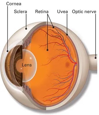
The extraocular muscles are attached to the white part of the eye called the sclera. This part of the eye is a strong layer of tissue covering nearly the entire surface of the eyeball.
The Surface of the Eye
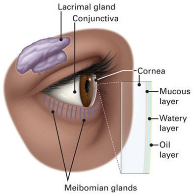
The eyes and inner surface of the eyelids are covered with a transparent membrane called the conjunctiva.
Tears lubricate the eye and makeup three layers. These three layers together are called the tear film. The conjunctiva makes the mucous layer. The lacrimal gland produces tears. The eye’s lacrimal gland sits under the outside edge of the eyebrow (away from the nose) in the orbit. The meibomian gland makes the oil that becomes another part of the tear film. Tears drain from the eye through the tear duct.
Front of the Eye
Light is focused into the eye through the clear, dome-shaped front portion of the eye called the cornea.
Behind the cornea is a fluid-filled space called the anterior chamber. The fluid is called aqueous humor. The eye is consistently producing aqueous humor. Aqueous humor also drains from the eye in the drainage angle area to maintain constant eye pressure.
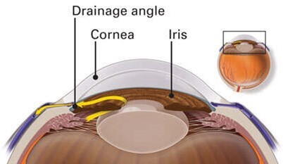
Behind the anterior chamber are the eye’s iris (the colored part of the eye) and the dark hole in the middle called the pupil. Muscles in the iris dilate (widen) or constrict (narrow) the pupil to control the amount of light reaching the back of the eye.
Directly behind the pupil sits the lens. The lens focuses light toward the back of the eye. The lens changes shape to help the eye focus on objects up close. Tiny fibers called zonules are attached to the capsule holding the lens, suspending it from the eyewall. The lens is surrounded by the lens capsule, which is left when the lens is removed during cataract surgery. Some types of replacement intraocular lenses go inside the capsule, where the natural lens once appeared.
By helping to focus light as it enters the eye, the cornea and the lens both play essential roles in giving us clear vision. 70% of the eye’s focusing power comes from the cornea and 30% from the lens.
Back of the Eye
The vitreous cavity lies between the lens and the back of the eye. A jelly-like substance called vitreous humor fills the hole.
The light focused into the eye by the cornea and lens passes through the vitreous to the retina; this is the light-sensitive tissue lining the back of the eye.
A tiny but specialized area of the retina called the macula gives us our detailed, central vision. The other part of the retina, the peripheral retina, provides us with our peripheral (side) vision.
The retina sends light as electrical impulses through the optic nerve to the brain. The optic nerve is made up of millions of nerve fibers that transmit these impulses to the visual cortex — the part of the brain responsible for our sight.
The Eye Organ Is Fascinating How It Works
The eye works just like a camera, where a camera’s shutter opens or closes depending on the amount of light needed to expose the film in the back of the camera. The eye connects to the brain and depends on the brain to interpret what we see. Our senses of sound, taste, hearing, and touch would not be completed without the sense of sight because sight is closely related to other parts of the anatomy. We hope this information is helpful for you, whether you are a patient seeking knowledge or simply curious about the wonders of the human body.

References: American Academy of Ophthalmology and the American Optometric Association. This blog provides information and discussion about eye health and related subjects. The content provided within this blog and any linked materials are not intended and should not be considered medical advice. If the reader or any person has a medical concern, they should consult with an appropriately licensed physician.